


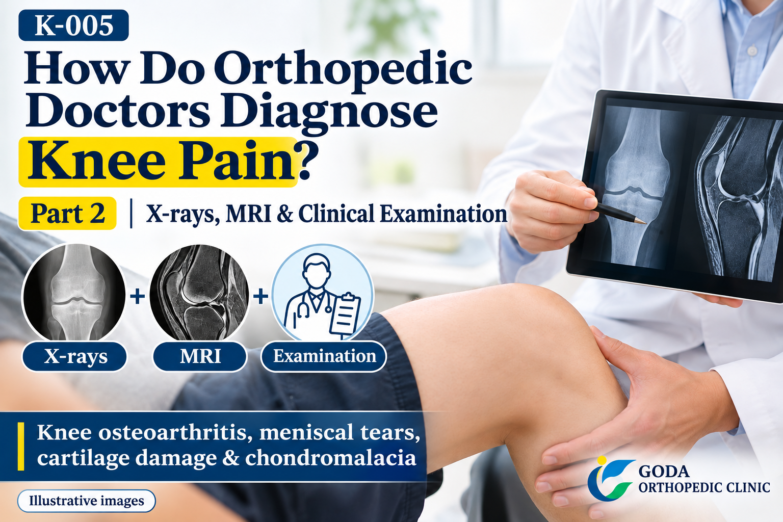
Part 2: How Do We Combine X-rays, MRI and a Clinical Examination to Identify the Cause?
Medical review
In Part 1, we explained how we assess walking, take a medical history, inspect and feel the knee, check its range of motion and areas of tenderness, and perform hands-on examination tests.
At Goda Orthopedics, we review the X-rays together with the patient only after completing this clinical examination.
Rather than looking at the images first and deciding, “There is a deformity here, so this must be the cause of the pain,” we check whether the imaging findings match the condition suggested by the examination.
14. We do not decide on a diagnosis by looking at the X-rays first
Patients with knee pain may already have had their X-rays taken before seeing the doctor.
Even then, the doctor first examines the patient.
After checking where the pain is, areas of tenderness, range of motion, instability and the results of hands-on tests, we look at the images together with the patient.
Imaging is very important, but it does not replace a clinical examination.
15. What do we look for on X-rays when assessing knee osteoarthritis?
On X-rays, we check for changes such as joint-space narrowing, osteophytes (bone spurs) and bone sclerosis (increased bone density), as well as alignment, including varus (bow-legged) and valgus (knock-kneed) alignment.
In knee osteoarthritis in particular, it is also important to ask, “Is too much of the load concentrated on the inner side?” and “How is the knee aligned overall?”
Articular cartilage itself cannot be seen directly on an X-ray.
As the cartilage becomes thinner, the joint space between the bones appears narrower. We use this finding to assess cartilage degeneration indirectly.
16. We compare the clinical examination findings with the X-rays
For example, the examination may reveal marked tenderness along the medial joint line, on the inner side of the knee.
The X-rays may also show narrowing of the medial joint space and varus deformity.
When the examination and imaging findings agree in this way, osteoarthritis affecting the inner compartment of the knee is more likely to be contributing to the pain.
On the other hand, if the changes on the X-rays do not adequately explain the symptoms, we consider other possible causes.

17. Knee pain may have more than one cause
Several conditions can be present in the same knee at the same time.
For example, osteoarthritis of the medial compartment, degeneration or a tear of the medial meniscus, damage to the articular cartilage on the inner side, and varus deformity may coexist.
If increasing deformity concentrates the load on the inner side, the medial joint space may narrow further, while laxity may develop on the outer side.
For some patients, we therefore cannot say, “This one condition is the only cause of your knee pain.”
Making a diagnosis is not simply a matter of naming one disease. It also means considering which of several findings are contributing to the current symptoms.
18. When we suspect a meniscal tear
Even if palpation, the McMurray test or a history of injury suggests a meniscal tear, the meniscus itself cannot be seen on X-rays.
In this situation, we consider MRI.
MRI can show tissues that plain X-rays cannot adequately assess, including the menisci, ligaments, cartilage, bone marrow and subchondral bone (the bone beneath the cartilage).
19. MRI is not needed for every patient with knee pain
MRI is a very useful examination, but not everyone with knee pain needs it.
If the medical history, clinical examination and X-rays adequately explain the cause, we may be able to start treatment without an MRI.
We consider MRI when we suspect a meniscal tear or ligament injury, want to assess cartilage damage, suspect a problem in the subchondral bone, cannot explain the cause from the X-rays, or find that the symptoms do not match the X-ray findings.
20. When a young person has pain despite no major abnormality on imaging
Older adults are more likely to have conditions that show up on imaging, such as knee osteoarthritis.
Younger people, especially those who play sports, may say, “My X-rays show no major abnormality, but my knee hurts when I exercise.”
In these cases, we also consider overuse, repetitive loading of the patellofemoral joint (where the kneecap meets the thighbone), cartilage damage, and problems with the tendons or surrounding tissues.
21. What is chondromalacia?
Chondromalacia patellae is one of the conditions we consider in young athletes and others with pain at the front of the knee.
In this condition, the articular cartilage on the back of the kneecap develops changes such as softening, cracks or degeneration.
Repetitive loading of the patellofemoral joint and abnormal tracking of the kneecap may be contributing factors.
Symptoms may include pain at the front of the knee when using stairs, squatting, running or sitting for a long time.
22. Chondromalacia and patellofemoral pain are not the same
This is an important medical distinction.
In the past, the term “chondromalacia” was sometimes used broadly for pain at the front of the knee.
Today, however, patellofemoral pain and chondromalacia, in which there are actual structural changes in the cartilage, are not considered to mean exactly the same thing.
Patellofemoral pain does not necessarily mean that cartilage damage is present.
Conversely, even if MRI or another examination shows cartilage changes, those changes may not be the sole cause of the pain.
23. Overuse is also important when diagnosing knee pain in young athletes
In young people, the MRI findings are not the only important information. We also need to know which sport they play, whether their training volume has increased, whether they repeat the same movement many times, whether rest improves the symptoms, and which specific movements cause pain.
Even without an obvious major injury, repetitive loading may cause symptoms around the patellofemoral joint or cartilage.
24. Does an abnormal MRI finding always explain the pain?
MRI provides detailed images, but a finding on an MRI is not necessarily the cause of the pain.
Degenerative changes in the menisci and other tissues may appear with age.
It is therefore important to assess the MRI by asking, “Do the images also show the condition suspected from the clinical examination?”
We do not make a diagnosis from the images in isolation from the patient.
25. Diagnosing a knee problem is not simply about naming one disease
For some patients, knee pain can be explained by one condition, such as a meniscal problem or knee osteoarthritis.
Other patients have knee osteoarthritis, meniscal degeneration, cartilage damage and alignment problems at the same time.
Making a diagnosis means considering which of these is contributing most to the current pain.
K-005 summary: Diagnosis means examining both the patient and the images
At Goda Orthopedics, our assessment of knee pain follows this sequence: a nurse takes the patient's history; we observe how the patient walks; the patient shows us where it hurts; we inspect the knee and feel around the kneecap; we assess range of motion, tenderness and instability; we perform hands-on tests such as the McMurray test; we compare the findings with the X-rays; and we use MRI when needed.
Imaging is important, but it cannot provide a diagnosis on its own. There are also things that a physical examination alone cannot tell us.
The most important part of diagnosing knee pain is to consider the patient's symptoms, clinical examination findings and images together.
Frequently asked questions about diagnosing knee pain
Q1. Can knee pain be diagnosed with X-rays alone?
X-rays can show changes in the shape of the joint and fractures, but some tissues, such as the menisci and ligaments, cannot be seen adequately on X-rays.
Q2. Would starting with an MRI give a more certain diagnosis?
Not necessarily. We first narrow down the possible causes through the medical history, clinical examination and X-rays, then perform an MRI if needed.
Q3. Does a positive McMurray test mean I have a meniscal tear?
It can raise suspicion, but it does not confirm the diagnosis on its own. We consider it alongside symptoms, tenderness, the history of injury and imaging findings.
Q4. What do you examine if my knee feels unstable?
We assess the collateral and cruciate ligaments using tests such as varus and valgus stress tests, anterior and posterior drawer tests, and the Lachman test.
Q5. If the X-rays show a deformity, is that the cause of my pain?
We check whether it matches the clinical examination findings. Several overlapping conditions may be contributing to the pain.
Q6. Can young people develop cartilage damage in the knee?
Yes. Sports and repetitive loading can cause cartilage damage, including damage to the cartilage of the kneecap.
Q7. What is chondromalacia?
It mainly refers to softening, cracks or degeneration in the articular cartilage on the back of the kneecap. However, not all pain at the front of the knee is chondromalacia.
Q8. Can knee pain have a cause that does not show up on imaging?
Yes. In young athletes in particular, overuse and other factors may contribute to pain even when imaging shows no clear major abnormality.
Related articles already published
- K-001: Where Does Your Knee Hurt? Possible Causes Based on the Location of Pain — Part 1
- K-002: When Does Your Knee Hurt? Possible Causes Based on the Movements That Hurt — Part 1: Walking, Stairs and Standing Up
- K-003: Symptoms That Accompany Knee Pain: Swelling, Fluid, Warmth, Catching and Giving Way
- K-004: Should You See a Doctor for Knee Pain? Pain You Can Monitor and Pain That Needs Prompt Assessment — Part 2: When to Monitor or See a Doctor
About the medical review
This article is based primarily on Dr. Goda's actual knee examination procedures and clinical experience in general orthopedic practice. It does not present a specialist opinion as a knee specialist.
The medical review for the original article checked the following points against standard medical knowledge: knee assessment systematically includes the medical history, inspection, palpation, range of motion and special tests; valgus stress tests assess the medial collateral ligament (MCL), while varus stress tests assess the lateral collateral ligament (LCL); the Lachman and anterior drawer tests are used to assess the anterior cruciate ligament (ACL), and the posterior drawer test to assess the posterior cruciate ligament (PCL); the McMurray test alone does not confirm a meniscal tear; MRI is useful for assessing the menisci, ligaments, cartilage and subchondral bone; and chondromalacia refers to structural changes in the cartilage of the kneecap and is not an exact synonym for patellofemoral pain.
Medical reviewer profiles
Chairman, GOZEN Medical Corporation; certified orthopedic specialist; Doctor of Medical Science.
Director, Kita 7-jo Goda Orthopedics; certified orthopedic specialist; Doctor of Medical Science; occupational physician; Japan Sport Association-certified sports doctor.
Director, Iwamizawa Goda Orthopedics; certified orthopedic specialist; Doctor of Medical Science; Japanese Society for Replacement Arthroplasty-certified physician; Japan Sport Association-certified sports doctor.
Article information
Article number: K-005
Article: How Do Orthopedic Doctors Diagnose Knee Pain? Medical History, Inspection, Palpation and Hands-on Tests
Part: Part 2: X-rays, MRI, overlapping conditions, knee pain in young people and chondromalacia
Series: Goda Orthopedics' Guide to Knee Pain
Medical review: Taketoshi Goda / Takuro Sasaki / Naoki Seito
Original Japanese publication date: October 25, 2026 (Sunday)
Original Japanese update date: October 29, 2026 (Thursday)
Related article: K-005 Part 1: Walking, Medical History, Inspection, Palpation and Hands-on Tests
This article was translated from the original Japanese using AI. Not all staff at Kita 7-jo Goda Orthopedics are fluent in English, but we make every effort to communicate with you and help you feel understood.


