



Looking at the menisci, ligaments, cartilage and inside the bones
Medical review: Taketoshi Goda, Takuro Sasaki and Naoki Seito
In Part 1, we explained why X-rays are usually the first imaging examination for knee pain.
However, some conditions cannot be identified with X-rays alone. This is where MRI and ultrasound come in. Each has different strengths.
11. The main purpose of MRI is to see what X-rays cannot show
A major purpose of knee MRI is to assess tissues that are difficult to see directly on X-rays.
These include the menisci, anterior cruciate ligament, posterior cruciate ligament, collateral ligaments, articular cartilage, bone marrow and subchondral bone (the bone beneath the cartilage).
The menisci and cruciate ligaments, in particular, cannot be seen directly on plain X-rays. MRI allows us to examine these structures inside the joint in detail.
12. MRI is important when a meniscal tear is suspected
We consider MRI when the history and clinical examination suggest a meniscal tear, based on findings such as tenderness along the joint line, pain with twisting, locking or the McMurray test.
With MRI, we can look at the actual images together with the patient and explain, “This is the meniscus,” and “There is a tear in this area.”
For young athletes, seeing what is happening inside their own knee can also help them understand their treatment and plan for a return to sport.
13. MRI can also assess ligament injuries
MRI is useful when we suspect an injury to the anterior cruciate ligament, posterior cruciate ligament or collateral ligaments, for example after a sports injury.
Of course, the diagnosis is not based on MRI alone. We consider the circumstances of the injury, feelings of instability, swelling and physical examination findings, including the Lachman test and anterior and posterior drawer tests, together with the MRI.
14. We also use MRI to assess cartilage
Articular cartilage cannot be seen directly on plain X-rays. MRI allows us to examine its condition in more detail.
For example, if a young athlete says, “I can walk normally, but it hurts when I exercise,” we may suspect a problem with the cartilage or around the patellofemoral joint, where the kneecap meets the thighbone.
MRI can sometimes help with the diagnosis of conditions such as chondromalacia, which we discussed in K-005.
15. MRI also looks inside the bones
MRI is useful for more than soft tissues. Its ability to show changes inside the bones is also very important.
For example, a subchondral insufficiency fracture of the knee may not show a clear abnormality on plain X-rays in its early stages. Even so, the patient may report sudden, severe pain, pain when putting weight on the knee or pain at night.
In these situations, we may use MRI to look for changes in the bone marrow or a fracture line.
If symptoms are severe despite normal X-rays, we need to consider a problem inside the bone.
16. “My X-rays are normal, but it really hurts” is important information
An important principle in diagnostic imaging is not to stop investigating simply because nothing appears on the X-rays.
If the examination suggests that the severity of the pain cannot be explained by the X-rays alone, we consider other possible causes involving the menisci, ligaments, cartilage or subchondral bone.
This is when MRI becomes important.
17. MRI helps us explain the condition, as well as diagnose it
Another reason for using MRI in clinical practice is to help explain the condition to the patient.
Especially for young athletes, looking at an MRI together and saying, “This is the meniscus, and this is where it is injured,” may be easier to understand than simply saying, “There is a problem with your meniscus.”
Images are not only a tool for the doctor to make a diagnosis. They also help patients understand their own condition.
18. Why not start with MRI?
Patients sometimes say, “If there are so many things X-rays cannot show, please do an MRI first.”
However, MRI alone does not assess everything. X-rays are particularly useful for examining bone shape, fractures, bone spurs, bone sclerosis, alignment and the joint space under weight-bearing conditions.
In particular, a standard MRI does not assess the joint space under the same conditions as an X-ray taken while standing with weight on the knee.
Rather than choosing MRI first simply because it is considered more advanced, it is important to choose the examination according to its purpose.
19. What can ultrasound show?
Knee ultrasound is another useful examination.
It is particularly suited to assessing joint effusion (fluid in the joint), synovitis (inflammation of the joint lining), Baker's cysts and superficial tissues such as the patellar tendon, quadriceps tendon, collateral ligaments, pes anserinus region on the inner side of the knee and iliotibial band.
Knee ultrasound can assess a wide range of superficial structures, including joint fluid, the synovium (joint lining), the extensor mechanism that straightens the knee, collateral ligaments, tendons, bursae and tissues at the back of the knee.
20. Ultrasound can show Baker's cysts clearly
Ultrasound is very useful when there is a Baker's cyst behind the knee. It can show the fluid-filled sac during the examination.
Ultrasound is a useful test for checking whether a Baker's cyst is present.
21. Ultrasound can also show parts of the menisci
Some parts of the menisci can be seen with ultrasound. However, MRI is more suitable for a detailed assessment of the whole meniscus.
Some areas are difficult to assess with ultrasound, particularly deep within the joint, the cruciate ligaments and the bone marrow.
Being able to see part of a meniscus on ultrasound does not mean that ultrasound provides the same information as MRI.
22. Ultrasound is not well suited to looking inside bones
Most ultrasound waves are reflected at the surface of the bone. Ultrasound is therefore not well suited to examining bone marrow, subchondral bone or abnormalities inside the bone in detail.
MRI is important when we suspect an internal bone abnormality, such as a subchondral insufficiency fracture.
23. Ultrasound may be used when MRI is difficult
Some patients find it difficult to undergo MRI. Possible reasons include severe claustrophobia, concerns identified during MRI safety screening or a physical condition that makes scanning difficult.
Metallic ingredients in some cosmetics or decorative items may also pose a problem, so safety screening at the MRI facility is necessary before the scan.
Having nail polish or nail art does not automatically mean you cannot have an MRI. You should check with the MRI facility in advance.
When MRI is difficult, ultrasound may be used as an additional assessment tool, depending on the condition.
24. Injections and aspiration can also be guided by ultrasound
Ultrasound-guided injection and aspiration involve advancing a needle while watching it on ultrasound.
Ultrasound guidance lets the clinician check the needle's position and is used to improve the accuracy of injections into the knee joint and aspiration of joint fluid.
At Goda Orthopedics, however, we often perform routine knee joint injections and aspiration using anatomical landmarks to guide needle placement.
There is no single method that is always the right one. The choice depends on the patient's condition, the purpose of the procedure, and the doctor's skills and experience.
25. How do we choose between X-rays, MRI and ultrasound?
Our basic approach to imaging for knee pain is as follows:
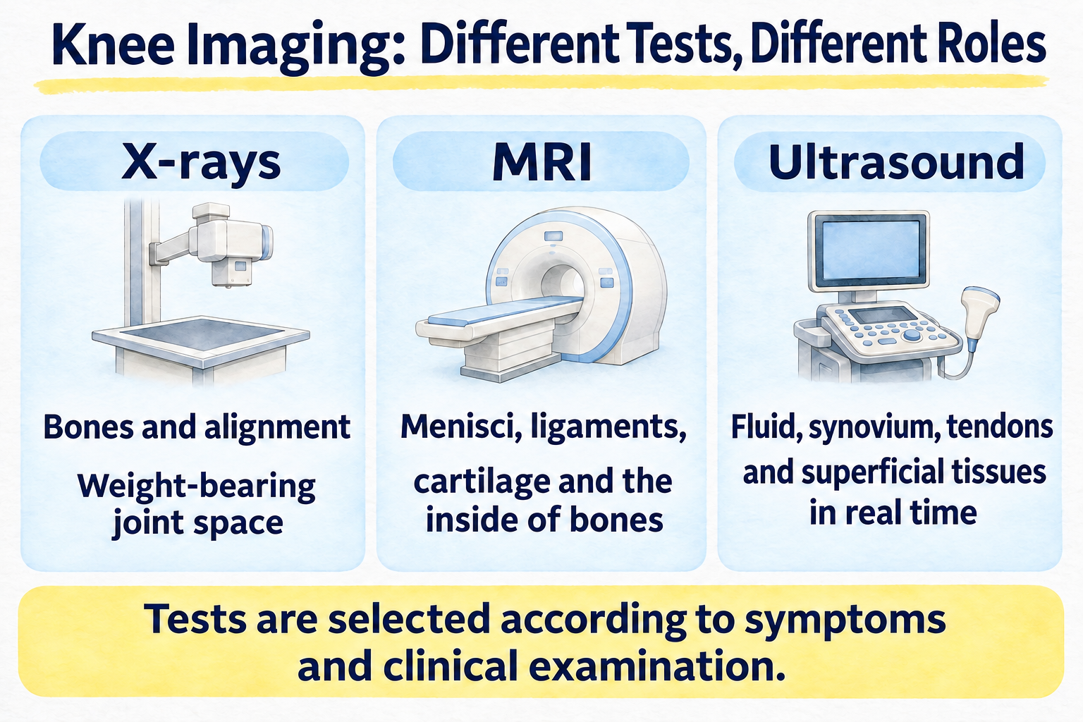
- X-rays: Examine bones, joint changes, alignment and the joint space under weight-bearing conditions.
- MRI: Examine the menisci, ligaments, cartilage, bone marrow and subchondral bone.
- Ultrasound: Examine joint effusion, the synovium, tendons, bursae and other superficial tissues in real time.
The key is to choose the examination based on the condition we suspect, rather than asking which test is best overall.
26. More tests do not necessarily mean a more accurate diagnosis
You may wonder, “Wouldn't it be reassuring to have every test?” However, adding tests that are unlikely to be needed does not necessarily improve the diagnosis.
We use the history and clinical examination to identify what we suspect, then choose tests to help answer that question.
At Goda Orthopedics, we select the tests each patient needs by following the sequence of clinical examination, X-rays, and then MRI or ultrasound if necessary.
K-006 summary: MRI is not simply “better” than X-rays
Each type of knee imaging has its own role.
- X-rays show bones, joint changes, alignment and the joint space under weight-bearing conditions.
- MRI examines the menisci, ligaments, cartilage and the inside of bones.
- Ultrasound is particularly useful for superficial problems such as joint effusion, synovitis, tendon conditions and Baker's cysts.
X-rays, MRI and ultrasound are not a ranking from one level to the next. They examine different things.
Most importantly, a diagnosis should bring together the patient's symptoms, clinical examination findings and imaging, rather than relying on test results alone.
Frequently asked questions about knee imaging
Q1. If my knee hurts, will starting with MRI give a clearer answer?
Not necessarily. X-rays provide important information about the bones, alignment and the joint space under weight-bearing conditions.
Q2. Can X-rays show cartilage?
Cartilage itself cannot be seen directly. Its condition is assessed indirectly through findings such as joint-space narrowing.
Q3. Can X-rays show the menisci?
The menisci cannot be seen directly on standard X-rays. MRI is considered when the clinical examination suggests a meniscal tear.
Q4. Can MRI find an abnormality even when X-rays are normal?
Yes. MRI may reveal abnormalities of the menisci, ligaments, cartilage or subchondral bone.
Q5. Why are standing X-rays taken for knee osteoarthritis?
They allow us to assess how much the joint space narrows when body weight is actually placed through the knee.
Q6. What can knee ultrasound show?
It is useful for assessing joint effusion, synovitis, Baker's cysts and superficial tissues such as the patellar tendon.
Q7. Can I have an MRI if I have nail polish or nail art?
These do not automatically prevent you from having an MRI. The approach depends on the product and the facility's safety standards, so please check with the MRI facility before your scan.
Q8. Are ultrasound-guided injections better?
Ultrasound guidance allows the clinician to check the needle's position, and studies have shown high accuracy. Injections using anatomical landmarks are also common for the knee joint. The method is chosen according to the patient's condition and the purpose of the procedure.
Published related articles
- K-001: Where Does Your Knee Hurt? Possible Causes Based on the Location of Pain — Part 1
- K-002: When Does Your Knee Hurt? Possible Causes Based on Painful Movements — Part 1: Walking, Stairs and Standing Up
- K-003: Symptoms That Accompany Knee Pain: Swelling, Fluid, Warmth, Catching and Giving Way
- K-004: Should I See a Doctor for This Knee Pain? When You Can Monitor It and When to Seek Care Soon — Part 2: When to Monitor or See a Doctor
- K-005: How Do Orthopedic Doctors Diagnose Knee Pain? History, Inspection, Palpation and Hands-on Examination Tests — Part 1 / Part 2
About the medical review
This article is based primarily on Dr. Goda's clinical experience and use of knee imaging in general orthopedic practice. It does not present a specialist opinion as a knee specialist or diagnostic radiologist.
The medical review for the original article checked the following points against standard medical knowledge: X-rays are the basic initial imaging examination for chronic knee pain; weight-bearing X-rays are useful for assessing joint-space narrowing in knee osteoarthritis; MRI is useful when X-rays do not adequately explain the cause; MRI is useful for assessing the menisci, ligaments, cartilage, bone marrow and subchondral bone; ultrasound is useful for assessing superficial problems involving joint fluid, the synovium, tendons and Baker's cysts; and individual safety screening is necessary before MRI, rather than automatically ruling out a scan simply because nail products or similar items are present.
Medical reviewers
Taketoshi Goda
Chairman, GOZEN Medical Corporation; certified orthopedic specialist; Doctor of Medical Science.
Takuro Sasaki
Director, Kita 7-jo Goda Orthopedics; occupational physician; Japan Sport Association-certified sports doctor.
Naoki Seito
Director, Iwamizawa Goda Orthopedics; certified orthopedic specialist; Doctor of Medical Science; Japanese Society for Replacement Arthroplasty-certified physician; Japan Sport Association-certified sports doctor.
Article information
Article number: K-006
Article: Tests for Knee Pain: What Can X-rays, MRI and Ultrasound Show?
Part: Part 2: MRI, ultrasound, menisci, ligaments, cartilage and the inside of bones
Series: Goda Orthopedics' Guide to Knee Pain
Medical review: Taketoshi Goda / Takuro Sasaki / Naoki Seito
Original Japanese publication date: October 29, 2026 (Thursday)
Original Japanese update date: Not yet determined
This article was translated from the original Japanese using AI. Not all staff at Kita 7-jo Goda Orthopedics are fluent in English, but we make every effort to communicate with you and help you feel understood.


