



Bones, joint changes, alignment and weight-bearing views
Medical review: Taketoshi Goda, Takuro Sasaki and Naoki Seito
“If MRI gives a more detailed picture, wouldn’t it be better to have an MRI first?”
Patients with knee pain sometimes ask us this question.
MRI is indeed an excellent examination for looking closely at structures that are difficult to assess on X-rays, including the menisci, ligaments, cartilage and the inside of bones.
However, MRI is not simply a higher-level version of an X-ray. X-rays and MRI look at different things.
At Goda Orthopedics, when we judge that imaging is needed for a patient with knee pain, we first take plain X-rays and compare them with the findings from the clinical examination. We then proceed to MRI if we suspect a condition that X-rays alone cannot reveal.
1. At Goda Orthopedics, we sometimes take X-rays before the doctor's examination
A nurse first asks about your symptoms and medical history. If this suggests that the main source of the pain is likely to be the knee joint, we may take X-rays before you enter the consultation room.
This helps reduce the burden of moving repeatedly between the consultation room and the X-ray room and makes the visit run more smoothly.
This does not mean that the doctor decides on a diagnosis by looking at the X-rays first. As explained in K-005, we first take your history, observe how you walk, inspect and feel the knee, check its range of motion and areas of tenderness, and perform hands-on examination tests. Only then do we review the X-rays together with you.
2. First, we look at the knee as a whole
When reading a knee X-ray, we do not start by focusing on just one part of the joint space. We first look at the whole knee.
One particularly important point is alignment. This means how the femur, knee joint and tibia line up: in simple terms, the overall alignment of the leg.
For example, we check for bow-legged alignment, knock-kneed alignment and differences between the two sides.
In knee osteoarthritis, this overall alignment matters as well as changes in the joint surfaces.
3. Next, we check the bones one by one
After looking at the knee as a whole, we examine each area, including the patella (kneecap), femur (thighbone), tibia (shinbone) and upper part of the fibula.
We check the bones in sequence for fractures, unusual shape, abnormal shadows within a bone and changes to the bone surface.
X-rays are a very important examination for looking at the bones themselves.
4. In knee osteoarthritis, we assess the joint space
The joint space is an important feature when assessing knee osteoarthritis. It is the gap seen between the femur and tibia on an X-ray.
This area normally contains articular cartilage and the menisci. However, cartilage itself does not appear directly on a standard X-ray.
We therefore assess cartilage wear and related changes indirectly by checking whether the joint space has narrowed.
In addition to joint-space narrowing, we look for X-ray findings of knee osteoarthritis such as osteophytes (bone spurs) and subchondral sclerosis (increased density in the bone beneath the cartilage).
5. We also check for bone spurs and bone sclerosis
As knee osteoarthritis progresses, various changes can occur in the bones. A typical example is an osteophyte, or bone spur: a projection of bone around the joint.
We may also see sclerosis, in which the bone beneath the cartilage appears denser.
We assess the state of knee osteoarthritis by considering these findings alongside the joint space, alignment, symptoms, areas of tenderness and range of motion.
6. Standing X-rays are important in knee osteoarthritis
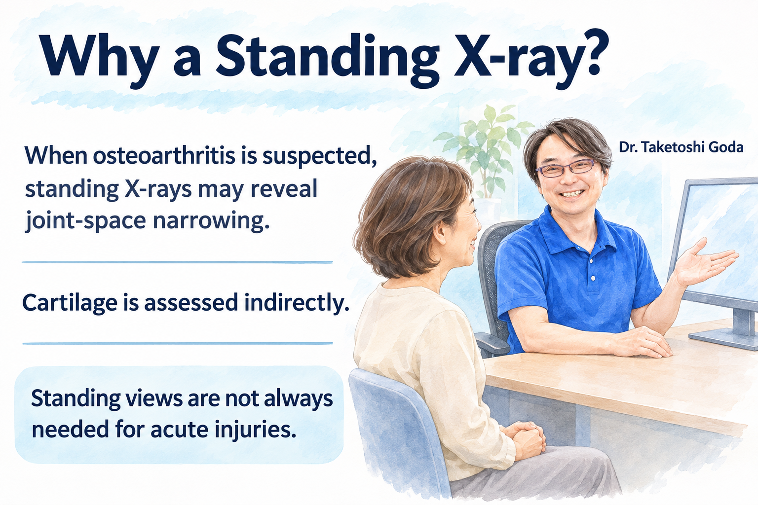
When knee osteoarthritis is suspected in a middle-aged or older adult, weight-bearing X-rays, taken while standing, can be important.
When you are lying down, your body weight is not loading the knee. When you stand, your weight is actually placed through it. This lets us see how much the joint space narrows under load.
Weight-bearing X-rays are useful for assessing joint-space narrowing in knee osteoarthritis.
7. X-rays taken lying down and standing can look different
For example, the joint space may appear reasonably well preserved when you are lying down but become clearly narrower when you stand and put weight on the knee.
In everyday life, patients experience pain during weight-bearing activities such as standing, walking and using stairs. This is why it is useful to assess an arthritic knee while it is actually bearing weight.
The appearance of the joint space also changes with the X-ray angle and the degree of knee bending, so we assess it from several directions when necessary.
8. Weight-bearing views are not always needed for acute injuries in younger people
When a younger patient injures their knee during sports or another activity, the purpose of the examination differs from an assessment for osteoarthritis.
In an acute injury, the main focus is on checking for fractures, abnormalities of the kneecap, abnormal bone alignment and dislocation.
There is no need to force a patient to have a standing, weight-bearing X-ray if the pain is so severe that putting the foot down and standing is difficult.
The examination method varies with the patient's age and condition.
9. Can X-rays show the menisci?
In general, the menisci themselves cannot be seen directly on plain X-rays. Similarly, standard X-rays alone cannot adequately assess the anterior cruciate ligament, posterior cruciate ligament, collateral ligaments or articular cartilage.
That does not make X-rays unhelpful. We first examine the bones, alignment and deformity. If symptoms remain that the X-rays cannot explain, we proceed to MRI or other examinations.
10. A normal X-ray does not mean there is no problem with the knee
This is a point we particularly want patients to understand.
Even if an X-ray shows no major abnormality, there may still be a meniscal tear, ligament injury, cartilage damage or an early lesion in the bone beneath the cartilage.
For chronic knee pain, if the initial X-rays are normal or show only fluid in the joint, MRI may be considered as the next examination.
Part 1 summary: X-rays have a different role from MRI
When assessing knee pain with X-rays, we examine the alignment of the whole knee; bones such as the patella, femur and tibia; the joint space; and changes such as bone spurs and bone sclerosis.
In knee osteoarthritis in particular, it is important to see how the joint space changes under weight-bearing conditions.
MRI can show some tissues in more detail. However, this does not mean that X-rays are unnecessary simply because MRI provides greater detail. X-rays and MRI examine different structures and serve different roles.
Continued in Part 2
Part 2 explains how MRI examines the menisci, ligaments, cartilage and the inside of bones; when MRI is needed; what ultrasound can show; alternatives when MRI is difficult; ultrasound-guided injections and aspiration; and how we choose between X-rays, MRI and ultrasound.
About the medical review
This article is based primarily on Dr. Goda's clinical experience and use of knee imaging in general orthopedic practice. It does not present a specialist opinion as a knee specialist or diagnostic radiologist. The descriptions of X-rays, weight-bearing imaging and imaging assessment of knee osteoarthritis have been checked against standard medical knowledge and edited for patients.
Medical reviewers
Taketoshi Goda
Chairman, GOZEN Medical Corporation; certified orthopedic specialist; Doctor of Medical Science.
Takuro Sasaki
Director, Kita 7-jo Goda Orthopedics; certified orthopedic specialist; Doctor of Medical Science; occupational physician; Japan Sport Association-certified sports doctor.
Naoki Seito
Director, Iwamizawa Goda Orthopedics; certified orthopedic specialist; Doctor of Medical Science; Japanese Society for Replacement Arthroplasty-certified physician; Japan Sport Association-certified sports doctor.
Article information
Article number: K-006
Article: Tests for Knee Pain: What Can X-rays, MRI and Ultrasound Show?
Part: Part 1: X-rays, bones, joint changes, alignment and weight-bearing views
Series: Goda Orthopedics' Guide to Knee Pain
Medical review: Taketoshi Goda / Takuro Sasaki / Naoki Seito
Original Japanese publication date: October 27, 2026
Original Japanese update date: October 29, 2026
Related article: K-005 Part 2: How Do We Combine X-rays, MRI and a Clinical Examination to Identify the Cause?
This article was translated from the original Japanese using AI. Not all staff at Kita 7-jo Goda Orthopedics are fluent in English, but we make every effort to communicate with you and help you feel understood.


